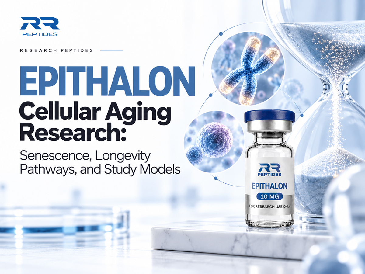
Why do some cells stop dividing while neighbouring cells remain active? The answer involves chromosome protection, stress signalling, metabolism, and decisions that determine whether a cell continues growing. Researchers investigate these complex processes through carefully selected laboratory models rather than treating aging as a single measurable event.
Epithalon cellular aging research examines a synthetic four-amino-acid peptide, also called epitalon, in relation to those biological questions. Early cell culture papers reported changes in telomerase activity and proliferative capacity. However, limited robust independent replication means those observations cannot establish rejuvenation, improved human health, or longer lifespan.
For Canadian laboratory teams, RR Peptides provides an Epithalon 10mg research listing and information about available material specifications. Explore the RR Peptides collection when planning a controlled investigation, while independently verifying documentation, suitability, and institutional requirements.
What Is Cellular Aging in Laboratory Research?
Aging Is a Network of Cellular Changes
Cellular aging describes changes that accumulate as cells divide, encounter stress, or adapt to their environment. Although these processes overlap, they do not develop at identical rates.
Common research areas include:
Genomic instability: Damage and imperfect repair can disrupt normal cellular regulation.
Telomere dysfunction: Unprotected chromosome ends may activate persistent checkpoint responses.
Mitochondrial alterations: Changes in energy metabolism can reshape stress signalling.
Proteostasis: Impaired protein folding and recycling may disturb cellular functions.
Epigenetic regulation: Gene activity can change without alterations to DNA sequence.
Cellular senescence: Some cells enter durable proliferation arrest with modified signalling.
Epithalon cellular aging research must identify which process an experiment measures. A change in one biomarker is not a comprehensive measure of biological age.
Replicative Senescence Versus Stress Responses
Replicative senescence can emerge after repeated division, particularly when telomeres become dysfunctional. By contrast, stress-induced senescence may develop after oxidative injury, oncogenic signalling, or other challenges without an extensive division history. Both states can involve stable growth arrest, although the pathways leading there may differ.
Senescence is also context dependent. It can restrict expansion of damaged cells and participate in tissue repair. Persistent senescent populations, however, may produce signalling molecules that alter surrounding tissue environments. Therefore, researchers should distinguish potentially protective arrest from harmful chronic accumulation.
Epithalon cellular aging research should assess proliferation, viability, and several senescence markers together instead of assuming every nondividing cell is senescent.
How Scientists Describe Culture Age
Passage number measures how frequently cells were transferred between vessels, not their exact replication history. Population doublings offer additional information, but calculations depend on accurate cell counts and losses. Epithalon cellular aging research must account for oxygen tension, nutrients, density, and handling because these factors influence aging-associated markers.
Measurement
What it indicates
Important limitation
Passage number
Culture handling history
Does not count divisions
Population doublings
Estimated replication history
Sensitive to cell loss
Telomere length
Chromosome-end repeat distribution
Cannot predict lifespan alone
Senescence markers
Features associated with arrest
No universal single marker
Cell viability
Proportion of living cells
Does not establish healthy function
Epithalon cellular aging research becomes more interpretable when studies document donor characteristics, medium composition, sampling intervals, and independent cultures.
Explore cellular aging pathways and telomerase research with RR Peptides’ research-grade Epithalon 10mg.
Why Is Epithalon Studied in Cellular Aging Models?
The Peptide and Its Research Background
Epithalon is a synthetic tetrapeptide with the amino-acid sequence Ala–Glu–Asp–Gly, frequently abbreviated AEDG. It has been investigated within a relatively small body of experimental literature on cellular regulation and aging-associated biology.
A 2003 laboratory publication reported telomerase catalytic-subunit expression, enzyme activity, and telomere elongation in cultured human fetal fibroblasts following experimental exposure. A subsequent paper described additional divisions in related cell models.
Epithalon cellular aging research therefore begins with a narrower question: can independent laboratories reproduce particular cellular observations under clearly defined conditions?
Questions Worth Testing
Useful experimental questions include:
Does a measured response exceed normal variability between matched cultures?
Are observed changes consistent across independent biological samples?
Do telomerase activity and telomere-length measurements agree?
Does altered proliferation reflect division, selection, or reduced cell death?
Are senescence markers consistent with a durable growth-arrest phenotype?
Do observations persist during follow-up after exposure ends?
Epithalon cellular aging research is more informative when each question corresponds to a predefined endpoint and suitable control groups.
Choosing an Appropriate Study Model
Different cell models answer different biological questions. Primary human fibroblasts are useful for investigating replication-associated changes, whereas transformed cell lines may already possess abnormal cell-cycle controls.
Model
Potential advantage
Key limitation
Primary fibroblasts
Replicative aging characteristics
Donor and passage variability
Immortalized lines
Convenient experimental consistency
Altered proliferation pathways
Stem cell cultures
Chromosome maintenance questions
Specialized baseline regulation
Stress-exposed cultures
Defined response pathways
Artificial conditions shape results
Animal tissues
Multicellular context
Species differences limit translation
Epithalon cellular aging research requires researchers to justify their chosen model and avoid generalizing findings from a single culture system.
Material Quality and Research Controls
Epithalon cellular aging research must consider peptide identity, purity, degradation, and production-batch differences when interpreting measurements. Analytical documents may help characterize materials, but a purity statement cannot establish biological efficacy.
Laboratories should consider identity confirmation, appropriate reference materials, storage records, and assay-compatible controls.
RR Peptides offers information about its Epithalon 10mg research material for teams investigating peptide-related questions. Explore available specifications and assess independent verification before selecting supplies for institutional laboratory work.
Epithalon cellular aging research depends on trustworthy material characterization as well as careful biological measurement.
Which Aging-Related Pathways Are Examined With Epithalon?
Telomerase and Chromosome Protection
Telomeres comprise repetitive DNA and protective proteins at chromosome ends. Telomerase can add telomeric repeats in certain cells, although activity varies by tissue and developmental state. Many cancer cells also depend on telomerase-associated maintenance, underscoring why enzyme activation is not automatically beneficial.
Early Epithalon publications linked experimental exposure with telomerase-related changes in specific cultured fibroblasts. Nevertheless, enzyme activity, telomere extension, cell growth, and organismal longevity are separate outcomes requiring separate evidence.
Epithalon cellular aging research should report telomerase activity alongside independently assessed telomere length and genomic stability rather than drawing conclusions from one assay.
Checkpoints That Control Cell Division
The p53–p21 and p16–RB pathways participate in growth arrest and cellular stress responses. Expression of their components may change during senescence, but individual markers are neither exclusive to senescence nor universally elevated in every model.
A convincing study would pair checkpoint measurements with proliferation assays, cell counts, and damage indicators. This approach helps distinguish stable arrest from temporary quiescence, nutrient deprivation, or toxicity.
Epithalon cellular aging research can examine checkpoint regulation as a hypothesis, without implying that the peptide has a confirmed direct effect on these pathways.
Mitochondria, Oxidative Stress, and Proteostasis
Mitochondria generate energy and participate in intracellular communication. Researchers may measure oxygen consumption, membrane potential, and oxidative-damage indicators. However, reactive oxygen species also serve normal signalling functions; simply reducing a signal does not necessarily represent improved cell health.
Protein-quality systems offer another perspective. Autophagy and protein degradation help cells recycle components, but static marker levels cannot reveal whether recycling is functioning efficiently. Functional assays and properly timed samples are necessary to interpret change.
Epithalon cellular aging research may include these measurements as exploratory outcomes; current evidence does not establish a consistent mitochondrial or proteostasis mechanism for Epithalon.
Inflammatory Signals and Cellular Communication
Some senescent cells release cytokines, growth factors, and other molecules grouped under the senescence-associated secretory phenotype, or SASP. Its composition varies by cell type, the stress that produced senescence, and the time elapsed since that event. A cytokine change alone does not prove a cell has become younger.
Potential measurements include:
Telomerase activity measured with validated enzymatic assays.
Telomere distributions assessed through complementary techniques.
Mitochondrial respiration evaluated with suitable normalization.
Protein turnover assessed through functional rather than static markers.
Secreted cytokines compared across matched cell populations.
Pathway endpoint
Possible observation
Unsupported conclusion
Telomerase activity
Enzymatic repeat addition
Longer human lifespan
Checkpoint expression
Altered cell-cycle signalling
Reversed human aging
Mitochondrial respiration
Bioenergetic differences
Proven clinical benefit
SASP measurements
Changed secreted factors
Improved immune function
Telomere distribution
Variation in chromosome ends
Overall biological age
Epithalon cellular aging research is stronger when investigators explain both what their data indicate and what the measurements cannot establish.
Explore cellular aging pathways and telomerase research with RR Peptides’ research-grade Epithalon 10mg.
How Is Cellular Senescence Evaluated in Epithalon Research?
Why One Biomarker Is Not Enough
Senescence is a complex cellular state rather than a diagnosis established by one stain. Senescence-associated beta-galactosidase activity is widely used, but positive staining can also arise under other conditions. Reduced DNA synthesis likewise may indicate temporary arrest, toxicity, or changes in culture conditions.
Epithalon cellular aging research should combine complementary markers with functional evidence and verify that apparently altered cells remain viable.
Measurement
Intended observation
Interpretation challenge
SA-beta-gal staining
Senescence-associated activity
Limited specificity
EdU incorporation
DNA synthesis
Low uptake has several causes
p16 and p21
Checkpoint expression
Context-dependent regulation
DNA-damage foci
Persistent damage responses
Not always telomere-specific
SASP profiling
Secreted signalling molecules
Timing and model sensitivity
Population doublings
Long-term proliferative behaviour
Accurate counts required
Epithalon cellular aging research benefits from agreement between marker patterns, growth history, and cell survival measurements.
Separating Senescence From Cell Death
A culture can contain fewer dividing cells because cells have died, differentiated, or temporarily paused division. Therefore, proliferation measurements should be interpreted with total cell counts, apoptosis indicators, viability tests, and microscopy when appropriate.
Controls should include matched untreated cultures, relevant vehicle comparisons, and independent cell preparations.
Epithalon cellular aging research should never label a reduction in proliferation as an anti-aging effect without ruling out plausible alternative explanations.
Building a Reproducible Experimental Design
Researchers should define their primary endpoint and analysis plan before collecting data. Biological replicates must represent independent samples, while technical replicates mainly describe measurement variability. Treating repeated wells from one culture as independent donors can exaggerate statistical confidence.
A practical reporting checklist includes:
Document cell origin, donor information, and passage history.
Describe peptide identification and relevant analytical records.
Identify primary outcomes, comparison groups, and sampling times.
Measure viability alongside proliferation and senescence endpoints.
Report biological sample numbers, effect sizes, and uncertainty.
Describe negative results, unexpected observations, and assay limitations.
Epithalon cellular aging research becomes more valuable when other laboratories can understand and independently reproduce the reported methods.
Tracking Responses Over Time
Longitudinal sampling helps determine whether an observation is temporary or persistent. Yet cultures change composition as some cells divide or survive more readily than others. A rise in average proliferative capacity could therefore reflect selective expansion rather than restoration of the same cells.
Sampling intervals should match the primary biological question. Early measurements may capture signalling changes, whereas later measurements can reveal durable arrest, population replacement, or instability. Researchers should report outcomes that remain unchanged as carefully as those that differ.
Epithalon cellular aging research should interpret time-dependent changes against matched controls and avoid equating temporary marker shifts with rejuvenation.
Explore cellular aging pathways and telomerase research with RR Peptides’ research-grade Epithalon 10mg.
What Questions Remain About Epithalon and Cellular Aging?
Independent Reproduction of Early Findings
The direct Epithalon literature is limited relative to the wider fields of senescence and telomere biology. Historical experimental reports warrant consideration, but findings need replication across independent laboratories, cell donors, and validated methods. A small number of positive studies cannot establish a broadly reliable effect.
Epithalon cellular aging research requires transparent reporting of unsuccessful experiments alongside results that support a proposed mechanism.
Identifying a Direct Molecular Mechanism
Epithalon cellular aging research cannot establish direct binding to a target from enzyme activity or gene expression alone. The observed effect could arise through indirect stress signalling, different cell survival rates, or assay interference. Mechanistic experiments must distinguish these possibilities before causal statements become justified.
Epithalon cellular aging research remains exploratory regarding direct molecular targets and how any proposed effects vary among cell types.
Translation Beyond the Laboratory
Human aging involves organs, immune regulation, metabolism, environmental exposures, and cancer surveillance. Cultured cells cannot reproduce those interactions. Even if a laboratory reproduces altered telomerase activity, that finding cannot demonstrate prevention of disease or increased human healthspan.
Studies examining prolonged proliferation must also monitor genomic stability. Extending cellular divisions is not necessarily beneficial when growth control is compromised.
Epithalon cellular aging research should make these distinctions explicit whenever discussing possible longevity implications.
Canadian Regulatory Considerations
Health Canada has identified epitalon among unauthorized injectable peptide drugs in public safety communications. A product described as laboratory research material is not thereby authorized for human administration, and research-only wording does not automatically exempt a product from applicable rules. Researchers must distinguish legitimate institutional investigations from consumer claims about anti-aging treatments.
Canadian teams should verify applicable requirements, ethics oversight, procurement policies, and material documentation before beginning regulated activities. Nothing about experimental availability establishes clinical safety or effectiveness.
What Better Studies Would Look Like
Unanswered question
Research priority
Are findings reproducible?
Independent laboratories and donors
Which models respond?
Comparisons across primary cell types
Is the mechanism direct?
Target-focused mechanistic experiments
Do changes persist?
Longitudinal follow-up assessments
Is stability maintained?
DNA damage and growth-control testing
Do assays agree?
Orthogonal measurement approaches
Epithalon cellular aging research should prioritize reproducibility and clearly defined endpoints over attention-grabbing statements about reversing time.
Epithalon is a synthetic tetrapeptide with the sequence Ala–Glu–Asp–Gly. It has been studied experimentally in selected contexts involving cellular regulation and telomerase-associated biology.
Does Epithalon Reverse Cellular Aging?
There is no reliable clinical evidence that Epithalon reverses aging in humans. Observations in cultured cells cannot establish organism-wide rejuvenation or longer lifespan.
Does Telomerase Activity Mean Longer Telomeres?
No. Enzyme activity and measured telomere length are distinct endpoints. Both require appropriate controls, and neither alone establishes improved health outcomes.
Is Cellular Senescence Always Harmful?
No. Senescence can restrict potentially dangerous proliferation and participate in normal tissue responses. Persistent accumulation may also contribute to unfavourable signalling in some contexts.
Which Cell Models Are Most Useful?
Primary human cultures can reveal aspects of replicative aging, while other models address specific mechanisms. Choice depends on the question and limitations of the system.
Is Epithalon Approved for Human Use in Canada?
A research listing does not establish Health Canada authorization. Canadian authorities have warned about unauthorized injectable products containing epitalon.
What Remains Uncertain?
Epithalon cellular aging research has not established a broadly replicated direct mechanism or clinically meaningful human longevity benefits. Independent investigations remain necessary.
Conclusion
Cellular aging involves overlapping changes in chromosome maintenance, metabolism, stress responses, and growth regulation. Reliable investigations interpret several endpoints together rather than treating isolated molecular measurements as proof of rejuvenation.
Epithalon cellular aging research has raised questions about telomerase and proliferative behaviour, but existing laboratory observations do not establish safe or effective anti-aging treatment for humans. Reproducible experiments and clear reporting remain essential.
For Canadian laboratory planning, explore the Epithalon 10mg research material information available from RR Peptides. Review specifications and independent documentation, and assess institutional and regulatory requirements before selecting experimental supplies.
Disclaimer: All products and compounds discussed are intended strictly for laboratory and research purposes. This article is provided for educational and informational purposes only. This article is not medical advice, therapeutic guidance, or instructions for human administration.
×
Subscribe & Get 10% Off!
Enter your email address below to subscribe to our newsletter and receive an instant 10% OFF discount coupon code sent directly to your inbox!
Thank you for subscribing! Your 10% discount coupon has been sent to your email. Please check your inbox!